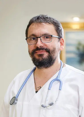
Dr. Razvan Ticulescu
Cardiologist at European Cardiomyopathy Center
Q&A: What is involved in cardiological follow-up of the HOCM patient after septal myectomy surgery?
Because septal myectomy surgery has a very good outcome, follow-up of the HOCM patient is fairly easy. It is one of the few operations after which the patient does not get a foreign material in the heart. A portion of the myocardium is removed, that which is responsible for the obstruction to the blood leaving the heart. The operation, like any heart surgery, can have certain complications. They are rare in our experience, over 100 patients operated on. We have not lost a single patient.
One of the lines we have to walk in following the patient is the electrical side, in the sense that in the heart there are some “wires” that conduct electric current. If you cut a portion of the muscle and remove that area, most often one of the wires that conduct the electrical current is also intricate. The others remain and generally no problems occur. One should watch and see how the other conduction pathways take over the transmission of the electrical impulse.
Another direction of follow-up: we have to follow the outcome of the intervention over time, which, according to our statistics and those from the international level, is very good. The removal of the obstruction, that gradient, is followed over time, sonographically. We cannot know if the operation has been successful or not except by doing an ultrasound. We do an ultrasound immediately in the operating theatre, then during hospitalisation and then we follow the patient periodically afterwards to see if this gradient/obstruction recurs. Generally, it does not recur. We have not had any patient who needed a new intervention because the obstruction recurred.
Another element that the technique used by our centre takes into account is the preservation of the mitral valve. During the operation, in addition to removing the muscle that prevents blood from leaving the heart, a repair of the mitral valve is performed, a mitral valve plasty is made.
The cardiologist is also responsible for monitoring the function of the mitral valve, in the sense that he or she will assess whether there is and how serious the mitral valve regurgitation is after this operation. What we have found is that the Ferrazzi technique used by our centre helps to reduce the mitral pathology associated with CMHO, so that mitral valve replacement is extremely rare. At this very moment, we are conducting a study in this regard and we have observed that even when there are important changes in the mitral valve, it is possible to repair and preserve the mitral valve, without having to replace it.
One thing that patients need to understand is that although the surgery will radically change their life in the sense that they will feel much better, they will not get tired, they will not have chest pain and their life expectancy will be normal, but the diagnosis of CMHO remains after the surgery. During the operation only a small portion of the myocardium is removed, the rest of the myocardium remains enlarged, it remains hypetrophic. This can lead to cardiac arrhythmias. For this reason, we monitor the heart rhythm through Holter examinations, which we repeatedly give to patients.
Also, also post-operatively, once the gradient/obstruction in the left ventricular ejection tract is dissipated, the risk of sudden death is reduced. This risk is assessed by the cardiologist according to several parameters: history of sudden death in the family, cardiac arrhythmias that still occur, presence of fibrosis that we evaluate by cardiac MRI. The cardiologist has to integrate all these data and it is not excluded to need a defibrillator implantation. Even if the gradient was eliminated and the patient was doing very well, however, the risk of cardiac arrhythmia and sudden death remained increased and a pacemaker may be needed.
Also, after surgery, drug treatment is required, which is much reduced compared to pre-op, but this beta-blocker treatment is important to be followed by the cardiologist.
After surgery, according to the existing protocol, the cardiologist should review the patient at one month, 3 months, 6 months and afterwards at one year, depending on each individual case. Patients who present certain problems are recommended to be re-evaluated more often.
Patient follow-up after surgery consists of: clinical examination, ultrasound, electrocardiogram, Holter monitoring and possibly cardiac MRI imaging.
